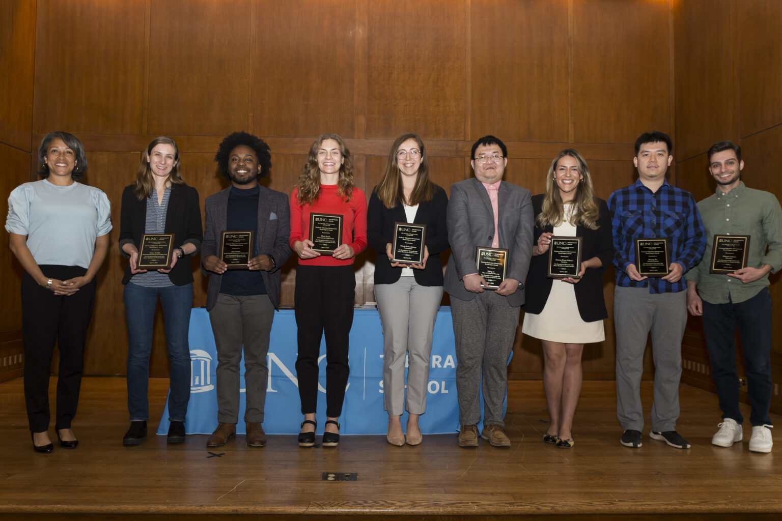
APS Doctoral Student Receives Impact Award for Research on Advanced Imaging Technology
Shuang Xu, a doctoral student in materials science in the department of applied physical sciences, is one of a dozen UNC graduate students who received the Graduate School’s Impact Award for his research on “Multisource Cone Beam Computed Tomography Using a Carbon Nanotube X-ray Source Array.”
Xu, who conducts his research as part of the Applied Nanotechnology Lab, received the award on April 4 at the George Watts Hill Alumni Center at the Graduate School’s 26th annual recognition celebration recognizing master’s degree and doctoral students. The Impact Award recognizes students whose research contributes to the educational, economic, physical, social or cultural well-being of North Carolina communities and citizens.
Xu is collaborating on the research with UNC-Chapel Hill Professors Christina Inscoe, Jianping Lu and Otto Zhou of the Department of Physics and Astronomy, Don Tyndall of the Adams School of Dentistry and Yueh Lee of the School of Medicine.
“I’m very happy about this,” said Xu. “This means that people think our research is valuable and impactful.”
Xu’s research focuses on multisource cone beam computed tomography (Ms-CBCT), a type of X-ray imaging, which has the potential to offer clinicians detailed 3-D imaging that will improve patient care and treatment. His research suggests that Ms-CBCT would be an improvement over conventional CBCT, which is a specialized medical imaging technique providing detailed 3-D volumetric images that offer a 360-degree spherical viewing angle of an object.
CBCT imaging is used primarily in the fields of dentistry, maxillofacial surgery, interventional radiology and image-guided radiation therapy, where precise visualization of hard-tissue structures like teeth, jaws and the skull is crucial. Despite its advantages, conventional CBCT has limitations, including reduced soft-tissue contrast, image distortion, the display of only hard tissues and hindered quantitative analysis.
Ms-CBCT overcomes these challenges by enhancing the accuracy of CT imaging of radiodensity of tissues by 60% and soft-tissue contrast by approximately 50%, allowing radiologists and clinicians to distinguish between different tissues and structures within the body. In addition to improving image quality and diagnostic accuracy, Ms-CBCT reduces the amount of “artifacts,” which can include the presence of metal objects, such as dental fillings or implants, and image distortion due to cone-beam imaging geometry.
“It’s a more comprehensive examination of the imaged area that can be particularly beneficial for capturing larger anatomical structures or regions of interest and improving soft-tissue contrast, making it possible to image more than just hard tissues,” said Xu.
When he isn’t conducting research, Xu, who hails from Jiangsu province in eastern China, said he likes playing video games and watching UNC and NBA basketball. Growing up, he and his friends rooted for their hero—7-foot, 5-inch-tall Yao Ming—who played center for the Houston Rockets. After getting a bachelor’s degree in physics, Xu migrated to the United States and enrolled at UNC-Chapel Hill because of the school’s outstanding reputation, especially in his field of study.
“The environment is very nice at UNC,” said Xu, who plans on pursuing a career in the medical imaging industry. “It’s a quiet small town, and it has introduced me to some of the best researchers in the world.”

Materials science Ph.D. student Shuang Xu, second from right, recently received the Graduate School's Impact Award.

